
How does image registration affect image-guided surgery accuracy?
In image-guided surgery, registration aligns preoperative or intraoperative images with the patient’s anatomy, establishing the spatial reference for navigation. When that alignment is inaccurate, the displayed position of an instrument may differ from its actual position. Even a small mismatch can matter when a target lies close to a critical structure or when the available surgical corridor is narrow.
For technical evaluators, the question is not simply whether a system reports a low registration error. It is whether the complete navigation process remains sufficiently accurate at the relevant anatomy, throughout the intended procedure, under conditions that resemble clinical use.
What registration does in image-guided surgery
A navigation system must relate several coordinate spaces: the image, the patient, the tracking system and the surgical instrument. Registration establishes a transformation between the image and the patient’s anatomy. Tracking then estimates the position of an instrument relative to that registered anatomy, allowing the system to display its apparent location on CT, MRI or other imaging data.
Consider a surgeon using a tracked pointer to identify a planned target on a preoperative CT scan. The navigation display can show the pointer at the target only if the image-to-patient registration, tracking geometry, reference attachment and pointer calibration are all sufficiently accurate. A convincing display is therefore not independent proof that the tip is where the screen says it is.
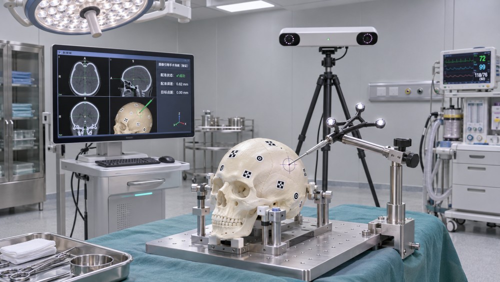
Registration is also not a one-time guarantee. The relationship established at the start of a procedure can become invalid if the patient moves relative to the reference marker, the anatomy shifts, or the image no longer represents the current surgical field. Some errors cause a broadly consistent offset; others vary across the operative region or appear only after a change in patient position.
Registration error is not the same as surgical accuracy
Suppliers may report several accuracy figures, but those figures answer different questions. Comparing them without their definitions can lead to an overly optimistic assessment.
- Fiducial registration error (FRE): the residual mismatch at the fiducial points used to calculate registration. A low FRE indicates that those points fit the calculated transformation well; it does not, by itself, establish accuracy at a separate surgical target.
- Target registration error (TRE): the discrepancy at a target point not used to compute the registration. TRE is often more relevant to navigation performance, although its value depends on target location, measurement method and test conditions.
- End-to-end navigation error: the discrepancy measured across a defined workflow, potentially including imaging, registration, tracking and instrument calibration. Its meaning depends on exactly which steps and sources of error were included.
- Clinical outcome: the result of the intervention, which also depends on planning, tissue behavior, surgical technique and other factors beyond navigation accuracy.
A single average can conceal occasional large deviations. Evaluators should request the distribution of errors, including the sample size, spread and worst observed values, rather than relying only on a mean or a best-case demonstration. They should also ask whether the measurement was taken near the intended target or only near the registration points.
Where inaccuracies enter the workflow
Image acquisition and segmentation
Registration cannot compensate reliably for anatomy that is poorly represented in the source image. Slice thickness, image artifacts, distortion, patient position and the visibility of landmarks can all influence the transformation. MRI distortion and CT artifacts, for example, may affect different regions in different ways. If a structure is segmented for planning or registration, the segmentation itself introduces another potential source of uncertainty.
The age of the image matters as well. An anatomically accurate scan obtained before surgery may no longer reflect the operative field after tissue is exposed, displaced or removed. Evaluators should distinguish image quality problems from changes that occur after acquisition: they require different controls.
Landmarks, surfaces and fiducials
Point-based registration uses corresponding landmarks or markers in image space and on the patient. Its performance depends on how accurately those points can be identified and localized. Poorly distributed points can yield acceptable residuals while leaving uncertainty at a distant target. Placing registration points around the region of interest, when clinically feasible, generally provides a more informative geometry than clustering them in one small area.
Surface-based registration matches sampled patient surfaces to an image-derived surface. It can reduce dependence on implanted markers, but performance is sensitive to the amount and distinctiveness of the sampled anatomy, soft-tissue deformation, occlusion and the quality of the image-derived surface. Automatic or hybrid methods still require validation: automation changes the workflow, not the need to verify the result.
Tracking, references and instruments
Optical tracking depends on visibility between cameras and markers. Electromagnetic tracking can operate without the same line-of-sight requirement, but its performance may be affected by the surrounding environment, including conductive or ferromagnetic materials. Neither approach should be judged solely by nominal tracker specifications; the installed operating-room configuration and instrument setup matter.
A reference frame must remain fixed relative to the anatomy being navigated. If a reference moves on the patient while the system continues to track it, the display may appear stable even though the image-to-anatomy relationship has changed. Instrument tip calibration, marker attachment and component integrity are equally important: a registration can be valid while the displayed tip position is wrong.
Anatomical change during surgery
Rigid registration assumes that the relevant relationship between anatomy and image can be represented without local deformation. That assumption can be reasonable for some bony structures, but less reliable for soft tissues. Brain shift, respiratory motion, changes in organ shape and tissue retraction can all reduce the correspondence between an earlier image and the current anatomy.
Intraoperative imaging or updated registration may help address selected changes, but neither is automatic proof of accuracy. The evaluator should ask what is updated, how the update is verified and whether the method is appropriate for the specific anatomy and procedure.
How should accuracy be verified during the procedure?
Registration quality should be checked at independent anatomical locations that were not used to compute the transformation. Ideally, these verification points are identifiable, clinically relevant and distributed near the intended working region. A visually plausible match at one convenient landmark is weaker evidence than consistent agreement across several suitable locations.
The operating team also needs a defined response when verification fails. Depending on the system and procedure, that may involve checking instrument calibration and reference fixation, repeating registration, acquiring updated images or using another clinically appropriate method. The system’s instructions for use and the institution’s procedures govern these decisions.
Reverification deserves attention after events that could disturb the navigation relationship: repositioning the patient, handling the reference frame, changing instruments, or observing a mismatch between the displayed position and visible anatomy. A warning or quality indicator is useful only if users understand what it measures and what it cannot detect.
Assessing supplier evidence: questions that reveal more than a specification
For procurement and clinical engineering teams, the most useful accuracy claim is one that can be traced to a defined test. Ask suppliers to provide the protocol and results for the intended system configuration, software version and clinical application—not merely a headline number.
Bench testing can be valuable for repeatability and comparison, but a controlled phantom does not reproduce every clinical variable. Conversely, clinical evidence may better represent actual workflow while making individual error sources harder to isolate. Both types of evidence should be interpreted in the context of the intended use.
Standards, documentation and acceptance planning
When reviewing a navigation system, request the manufacturer’s applicable regulatory documentation, risk management information and instructions for use for the relevant market and configuration. Standards may support aspects of medical device safety, software lifecycle processes, usability and quality management, but compliance with a general standard should not be mistaken for a guarantee of registration accuracy in a particular procedure.
Ask which standards and editions the manufacturer applies, how conformity is demonstrated, and whether the evidence covers the accessories and software being purchased. Particular attention should be paid to stated indications, contraindications, operating conditions, known limitations and required user training. Claims outside the documented intended use need separate scrutiny.
Before installation, agree on an acceptance plan with the clinical team and supplier. It should identify the configuration to be tested, the available test tools, responsibilities, pass criteria derived from the intended use and manufacturer’s documentation, and the process for recording failures. Routine checks should also be practical enough to perform within the real operating-room schedule.
The practical test: confidence at the surgical target
Image registration affects image-guided surgery accuracy by determining how faithfully the navigation display represents the patient’s anatomy. Yet registration is only one part of an error chain that includes imaging, tracking, reference stability, instrument calibration and anatomical change.
The strongest evaluation therefore combines independent target verification, transparent test data and a workflow for recognizing when navigation can no longer be trusted. For technical evaluators, the goal is not to find a single reassuring number; it is to understand where the system performs well, where uncertainty remains and how the clinical team is expected to manage it.
Have questions about the global trade trends discussed here, or planning your expansion and supply chain strategy? Leave a message below and let the trade experts at MTHH guide you.
We respect your privacy. Your information is strictly confidential.
Related News
- How to assess a clinical engineering equipment supplier's maintenance supportNov 22, 2026Assess a clinical engineering equipment supplier with confidence. Compare response times, maintenance plans, spare parts, and contracts to reduce downtime.
- How does equipment condition affect medical imaging equipment prices?Nov 21, 2026Medical imaging price depends on equipment condition. Compare new, refurbished, and used systems, uncover hidden costs, and make a smarter buying decision.
- How healthcare infrastructure in Saudi Arabia shapes demand for medical imaging equipmentNov 18, 2026Explore how healthcare infrastructure Saudi Arabia influences medical imaging demand, from hospital expansion to procurement and investment opportunities.
- How does image registration affect image-guided surgery accuracy?Nov 15, 2026Image-guided surgery accuracy depends on reliable registration. Explore error sources, verification methods and key questions to ask suppliers.