
How to compare magnetic resonance imaging systems for hospitals by capacity
Start with capacity, not magnet strength
Hospitals comparing MRI systems often begin with a familiar question: 1.5T or 3T? Field strength matters, but it does not determine whether a scanner can absorb the hospital's workload, protect access for urgent patients, or produce a financially sustainable imaging service. Capacity is the more useful starting point because it connects the MRI system to the work the hospital actually needs to perform.
For technical evaluation teams, “capacity” should mean more than the number of examinations expected each day. It includes the mix of examinations, time per slot, repeat-scan risk, patient preparation, contrast workflow, inpatient transport, anesthesia support, reporting turnaround, planned downtime, and the ability to recover after disruptions. A scanner that performs well on a standard outpatient brain protocol may still become a bottleneck when the same department must accommodate complex spine studies, cardiac imaging, sedated children, emergency cases, and hospitalized patients.
The practical comparison is therefore not between isolated specifications. It is between MRI configurations that can reliably deliver a required number of clinically acceptable examinations within the hospital's real operating conditions. A lower-priced system can be the better choice for a modest and predictable caseload. A more capable system may be justified where patient throughput, protocol complexity, or specialist service lines would otherwise create delays that the department cannot manage.
Define the workload the scanner must carry
Before reviewing vendor proposals, the evaluation team should convert demand into a workload profile. Monthly examination totals are useful for budgeting, but they can conceal the factors that determine daily pressure on the MRI room. A hospital with a moderate annual volume and a large share of long neurological, musculoskeletal, oncology, or cardiac protocols may need more scan capacity than a facility with higher volume but a narrow range of routine studies.
A useful profile separates patients and protocols into groups with different operational consequences:
- Routine outpatient examinations with relatively predictable preparation and scan times.
- Complex protocols requiring multiple anatomical regions, contrast phases, advanced sequences, or post-processing.
- Emergency and inpatient cases, where patient transport, monitoring, mobility limitations, and clinical instability affect room turnover.
- Pediatric, claustrophobic, bariatric, or pain-limited patients who may require extra preparation, wider bore access, immobilization, sedation, or repeat sequences.
- Specialist examinations such as cardiac, breast, prostate, liver, vascular, neurofunctional, or orthopedic imaging, depending on the hospital's service scope.
This exercise should produce a realistic schedule model rather than an optimistic count of scans per hour. Time must be allocated for screening, changing, IV access where required, coil placement, patient positioning, contrast administration, cleaning, image review, and recovery from delayed appointments. If the hospital plans to reserve slots for emergency demand, that reserve should be explicit. Treating every available minute as bookable outpatient time is a common source of capacity overstatement.
Technical teams should also ask how demand will be distributed. A single MRI unit can serve a planned outpatient list effectively when referrals are stable and urgent access is limited. It becomes more difficult when inpatient cases are inserted throughout the day or when emergency physicians expect rapid availability. In those environments, a high-throughput configuration, extended operating hours, or a second unit may be more relevant than purchasing a scanner with impressive peak sequence performance.
Compare throughput as a workflow capability
Vendor discussions often use scan time as shorthand for throughput. It is an important metric, but it is incomplete. The relevant measure is completed, diagnostically acceptable examinations per operating session, including the operational time around scanning. A protocol that is technically fast but produces frequent motion artifacts, difficult coil handling, or inconsistent positioning can reduce capacity rather than increase it.
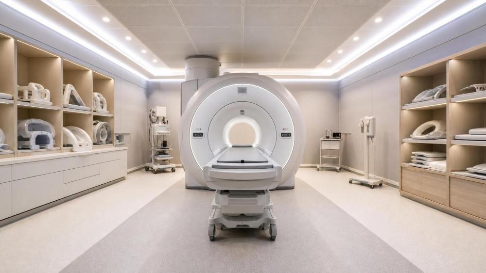
When evaluating magnetic resonance imaging systems for hospitals, request protocol-level timing assumptions for the examinations the department performs most often. The comparison should state what is included in each estimate: acquisition time only, table time, contrast workflow, planning scans, reconstruction, or total room occupancy. Without that distinction, two proposals can appear comparable while describing different parts of the patient journey.
Automation deserves careful scrutiny. Automated positioning, protocol selection support, standardized exam cards, acceleration techniques, and reconstruction tools can shorten routine workflow and reduce variation between operators. Their value depends on implementation quality. A hospital should ask to see how these functions work within its own protocol set, including difficult patients and common exceptions. A demonstration based only on idealized cases does not show whether the system will improve a busy clinical schedule.
Match clinical scope to field strength and configuration
A 1.5T MRI remains a broadly capable platform for many hospital imaging services. It can support routine neurological, musculoskeletal, body, vascular, and oncology applications when equipped and configured appropriately. For facilities building or replacing a general diagnostic service, the decision often turns on uptime, patient access, image consistency, coil coverage, workflow, and service support more than on pursuing the highest field strength available.
A 3T system may offer meaningful clinical and operational advantages for selected applications, particularly where higher signal performance can support finer detail, advanced neuroimaging, certain musculoskeletal examinations, prostate imaging, spectroscopy, or research-oriented work. However, it can also bring greater sensitivity to artifacts, protocol optimization demands, and differences in implant-related imaging considerations. It should be selected because the hospital has an established or planned clinical need, suitable radiologist expertise, and enough relevant demand to use the capability consistently.
There is no universal rule that higher field strength equals higher departmental capacity. If a hospital's caseload is dominated by routine general imaging and its MRI staff must manage a varied patient population with limited time for protocol development, a well-supported 1.5T system may provide more dependable output. Conversely, a specialist center may find that a 3T platform reduces scan time for certain protocols or supports referrals that a general configuration cannot address. The evaluation should identify which protocols would materially change and whether that change affects diagnostic confidence, schedule performance, or referral patterns.
Configuration choices can matter as much as field strength. Coil portfolio, gradient performance, patient table design, respiratory and cardiac gating tools, reconstruction capability, in-room communication, audiovisual patient support, and integration with contrast injection or monitoring systems all influence practical use. A scanner should be assessed as a complete clinical system, not as a magnet with a list of optional features.
Do not treat room infrastructure as a separate project
MRI capacity can be compromised before the first patient is scanned if room planning is handled after system selection. Site constraints affect the range of equipment that can be installed, the installation schedule, patient flow, safety zoning, and future service access. Technical evaluators should involve facilities, clinical engineering, radiology operations, infection-control stakeholders where applicable, and the selected vendor early enough to resolve these issues before procurement commitments become difficult to change.
The review should cover structural loading, equipment route, room dimensions, radiofrequency shielding, magnetic shielding requirements, electrical supply, cooling, quench arrangements, ventilation, fire and emergency planning, access control, and the location of the equipment room and control room. Hospitals replacing an existing MRI should verify whether legacy infrastructure remains suitable for the proposed system; assumptions based on the installed scanner can be costly when a new design has different spatial, electrical, or cooling needs.
Patient flow is equally important. The route for outpatient patients, inpatients on beds, emergency arrivals, bariatric patients, and sedated patients should be mapped through screening, changing, preparation, scanning, and recovery. A room layout that appears adequate on a technical drawing can produce slow turnover if staff must repeatedly move equipment, wait for elevators, or share constrained preparation areas with other services.
MRI safety planning belongs in the same review. The hospital needs operationally workable controls for screening, access to the magnet environment, emergency response, ferromagnetic risk management, and the use of MRI-compatible equipment. A system selected for high capacity will not achieve it if safety procedures are improvised, poorly understood, or incompatible with the patient groups the service intends to accept.
Test lifecycle assumptions before comparing price
Purchase price is only one element of MRI cost. A capacity-based comparison should consider the cost of operating the scanner over its intended service life, including installation work, energy and cooling demand, cryogen-related considerations where relevant, service coverage, planned maintenance, software upgrades, coils and accessories, training, downtime exposure, and eventual technology refresh.
Service terms deserve the same technical attention as image quality. Teams should establish what the service agreement covers, how remote diagnostics are used, how faults are prioritized, what response and restoration commitments are offered, whether critical components are regionally available, and whether preventive maintenance must occur during normal operating hours. Contract language should distinguish a response from a return-to-service outcome. A rapid acknowledgement of a fault does not restore lost scanning capacity.
It is also important to examine the hospital's own operating model. A scanner with advanced features may require more intensive applications training, protocol optimization, or specialist support. Those costs can be justified when the hospital intends to build complex service lines, but they should not be hidden inside a generic capital comparison. The same applies to software packages: evaluate which applications will be used in the first years of operation, which require future licenses, and who will maintain protocols as clinical demand changes.
Use an evaluation matrix that exposes trade-offs
A weighted scorecard can help multidisciplinary teams compare proposals, provided it does not turn clinical decisions into a purely numerical exercise. The scoring criteria should reflect the hospital's workload profile and should separate mandatory requirements from desirable features. A vendor should first demonstrate that it meets safety, site, service, interoperability, and core clinical requirements. Only then should comparative scoring determine which configuration best fits the institution's priorities.
For a general hospital, the highest weights may sit with routine throughput, patient accommodation, broad clinical coverage, service continuity, and infrastructure fit. A tertiary or specialist center may assign greater weight to advanced applications, high-end coil coverage, research compatibility, or complex protocol performance. The criteria should be agreed before demonstrations and commercial negotiations, otherwise optional features can distort the choice late in the process.
The final decision should read as a clear operational argument: this MRI system can deliver the required examination mix, within the available room and staffing model, with acceptable clinical performance and a credible plan for uptime and lifecycle support. That is a stronger basis for selection than choosing the scanner with the longest specification sheet or the lowest initial quotation.
Have questions about the global trade trends discussed here, or planning your expansion and supply chain strategy? Leave a message below and let the trade experts at MTHH guide you.
We respect your privacy. Your information is strictly confidential.
Related News
- How to choose diagnostic imaging equipment for small clinicsNov 14, 2026Choose diagnostic imaging equipment for clinics with confidence. Compare clinical needs, costs, space, workflows, and supplier support before you buy.
- How to size medical gas pipelines for peak demandNov 09, 2026Medical gas pipeline systems design starts with peak demand. Learn how to size pipes, calculate pressure loss, and check critical paths for reliable performance.
- What Trendelenburg angle should an operating table provide?Nov 05, 2026Compare operating tables Trendelenburg angles, load limits and accessories. Discover what to check before choosing the right table for your procedures.
- How to estimate the total cost of hospital imaging equipmentNov 04, 2026Plan hospital capital equipment imaging investments with confidence. Learn to estimate installation, service and operating costs and compare bids fairly.